Module: Overview
🧬 Virginia Biology SOL

DNA & Protein Synthesis

A self-paced lesson aligned to Virginia Biology SOL BIO.3 β€” exploring how DNA stores genetic information and directs protein synthesis.

🎯
SOL BIO.3 Focus: The student will investigate and understand that DNA has structure and is the foundation for protein synthesis, and that the structural model of DNA has developed over time.
πŸ“‹ What You Will Learn
  • How scientists discovered that DNA is the genetic material
  • How the double helix model of DNA was developed
  • The structure of DNA β€” nucleotides, base pairing, and the double helix
  • How DNA replicates (copies itself) before cell division
  • The differences between DNA and RNA
  • The three types of RNA: mRNA, tRNA, and rRNA
  • How transcription converts DNA β†’ mRNA
  • How translation converts mRNA β†’ protein
πŸ“š
8 Modules
Self-paced content
βš—οΈ
Virtual Lab
DNA Builder & Codon Decoder
πŸ“
SOL Quiz
10 SOL-style questions
πŸ“– Vocabulary

Key Terms

Learn these terms β€” they appear on the Virginia Biology SOL and are essential for understanding DNA and protein synthesis.

DNA
Deoxyribonucleic Acid β€” the molecule that carries genetic instructions for the growth, development, and function of all living things.
Core
Nucleotide
The building block (monomer) of DNA and RNA. Each nucleotide has three parts: a sugar, a phosphate group, and a nitrogenous base.
Structure
Double Helix
The twisted ladder shape of DNA. Two strands of nucleotides are held together by hydrogen bonds between base pairs.
Structure
Base Pairing
The rule that A pairs with T, and G pairs with C in DNA. In RNA, A pairs with U. These are held together by hydrogen bonds.
Structure
Complementary Strand
A strand of DNA or RNA whose base sequence matches and pairs with another strand following base pairing rules.
Structure
DNA Replication
The process by which DNA makes an exact copy of itself before a cell divides, so each new cell gets complete genetic information.
Process
RNA
Ribonucleic Acid β€” a single-stranded molecule that carries information from DNA and helps build proteins. Contains uracil (U) instead of thymine.
RNA
mRNA
Messenger RNA β€” copies the DNA code and carries it from the nucleus to the ribosome where proteins are made.
RNA
tRNA
Transfer RNA β€” carries amino acids to the ribosome and matches them to the correct codon on the mRNA during translation.
RNA
rRNA
Ribosomal RNA β€” makes up the ribosome structure. The ribosome is where proteins are assembled during translation.
RNA
Transcription
The first step of protein synthesis β€” DNA is used as a template to make mRNA. Happens in the nucleus.
Process
Translation
The second step of protein synthesis β€” the ribosome reads the mRNA and uses tRNA to build a chain of amino acids (a protein).
Process
Codon
A group of three mRNA bases that codes for one specific amino acid. AUG is the start codon; UAA, UAG, UGA are stop codons.
Genetic Code
Amino Acid
The building blocks of proteins. Each codon on mRNA codes for one amino acid. Amino acids are linked together to form a protein chain.
Protein
RNA Polymerase
The enzyme that reads the DNA template and builds the mRNA strand during transcription.
Enzyme
Protein Synthesis
The two-step process (transcription + translation) by which cells use genetic information in DNA to make proteins.
Process
πŸ”¬ Module 1

Discovery of DNA

How did scientists figure out that DNA β€” not protein β€” is the molecule that carries genetic information? It took several key experiments over many decades.

🎯
SOL Connection: The structural model of DNA has developed over time through scientific investigation.
SLIDE 01 / 05
🧫

Griffith's Experiment (1928)

Frederick Griffith studied two strains of bacteria β€” a harmless strain (R) and a deadly strain (S) that killed mice.

He killed the deadly S bacteria with heat, then mixed the dead S with live R bacteria. Surprisingly, the mice still died.

Griffith concluded that a "transforming principle" had passed from the dead S bacteria into the live R bacteria, making them deadly. He did not know what that substance was.
SLIDE 02 / 05
πŸ”¬

Avery, MacLeod & McCarty (1944)

Picking up from Griffith, this team systematically destroyed different molecules β€” proteins, RNA, then DNA β€” to find what the "transforming principle" was.

When they destroyed DNA, the transformation stopped. When they destroyed proteins or RNA, transformation still occurred.

βœ… Conclusion: DNA β€” not protein β€” is the molecule that carries genetic information. This was the first strong scientific evidence, though many scientists remained skeptical.
SLIDE 03 / 05
πŸ§ͺ

Hershey & Chase Experiment (1952)

Alfred Hershey and Martha Chase used viruses (bacteriophages) that infect bacteria. They labeled the virus's DNA with radioactive phosphorus (³²P) and its protein with radioactive sulfur (³⁡S).

After the virus infected bacteria, they found ³²P (DNA) inside the bacteria and ³⁡S (protein) outside.

βœ… Conclusion: The virus injects its DNA β€” not protein β€” into the host cell. DNA is the genetic material. This was definitive proof.
SLIDE 04 / 05
πŸ“

Chargaff's Rules (1950)

Erwin Chargaff analyzed DNA from many different organisms and found a consistent pattern:

πŸ”΄ Amount of Adenine (A) always = amount of Thymine (T)
🟒 Amount of Guanine (G) always = amount of Cytosine (C)

This became known as Chargaff's Rules. Although he wasn't sure why, these ratios strongly hinted that A-T and G-C were paired together in the molecule β€” a clue Watson and Crick used to build the correct model.
SLIDE 05 / 05
πŸ“Έ

Photo 51 β€” Rosalind Franklin (1952)

Rosalind Franklin used a technique called X-ray crystallography to study the structure of DNA. She fired X-rays at crystallized DNA and captured the diffraction pattern.

Her image β€” Photo 51 β€” revealed:
β€’ DNA has a helical (spiral) shape
β€’ The helix repeats every 3.4 Γ… (10 base pairs per turn)
β€’ The width of the helix is 20 Γ…

This data was shared with Watson and Crick without Franklin's knowledge and was essential for building the correct double helix model.
πŸ“… Key Experiments Timeline
1928
Griffith's Experiment
Frederick Griffith showed that something from dead, disease-causing bacteria could permanently transform harmless bacteria into dangerous ones. He called this the "transforming principle" β€” but did not know what it was.
1944
Avery, MacLeod & McCarty
Using Griffith's experiment, this team identified the "transforming principle" as DNA. This was the first strong scientific evidence that DNA β€” not protein β€” carries genetic information.
1950
Chargaff's Rules
Erwin Chargaff discovered that in any sample of DNA, the amount of Adenine always equals Thymine, and the amount of Guanine always equals Cytosine. This hinted at a paired structure.
1952
Hershey & Chase Experiment
Using viruses to infect bacteria, Hershey and Chase proved that DNA β€” not protein β€” is injected into cells and acts as genetic material. This was the definitive proof the scientific community needed.
1952
Photo 51 β€” Rosalind Franklin
Rosalind Franklin used X-ray crystallography to capture Photo 51 β€” an image showing DNA's helical shape and key measurements. Her data was essential to building the correct model.
1953
Watson & Crick's Double Helix Model
James Watson and Francis Crick used Franklin's data and Chargaff's rules to build the correct model of DNA β€” a double helix. Published in the journal Nature, April 1953. They received the Nobel Prize in 1962.
πŸ§ͺ Module 2

Models of DNA

Science improves over time as new evidence is gathered. Before Watson and Crick's accepted model, other scientists proposed different structures for DNA.

🎯
SOL Connection: The structural model of DNA has developed over time β€” science is a process of testing and revising ideas with evidence.
SLIDE 01 / 03
🧬

Early Ideas: Tetranucleotide Model

Phoebus Levene (1910s–1930s) believed DNA was a simple, repeating unit of the four bases in equal amounts. He thought it was too boring to carry genetic information β€” a conclusion that later turned out to be wrong.
SLIDE 02 / 03
πŸ“Έ

Franklin's X-ray Evidence

Rosalind Franklin's Photo 51 (1952) gave crucial evidence: DNA is a helix, the distance between base pairs is 3.4 Γ…, and the helix is 20 Γ… wide. This experimental data made it possible to build the correct model β€” yet her contribution was not fully recognized at the time.
SLIDE 03 / 03
πŸ†

Watson & Crick's Double Helix (1953)

Combining Franklin's X-ray data and Chargaff's base-pairing rules, Watson and Crick built the accepted double helix model: two antiparallel strands with bases pointing inward (A–T, G–C) and the sugar-phosphate backbone on the outside. This model correctly predicted how DNA could be copied.
πŸ’‘ Science works by building on evidence. Watson and Crick succeeded because they combined evidence from multiple sources β€” X-ray data, base-pairing rules, and chemical knowledge β€” rather than working from assumptions alone.
πŸ”© Module 3

DNA Structure

DNA looks like a twisted ladder. Understanding its structure explains how it stores information and copies itself.

🎯
SOL Connection: DNA is a double-stranded molecule made of nucleotides. Free nucleotides bond to the template using base-pairing rules (A–T and G–C).
SLIDE 01 / 05
🧱

The Building Block: A Nucleotide

DNA is a polymer β€” a long chain of repeating units. Each unit is called a nucleotide. Every nucleotide has three parts:

1️⃣ A deoxyribose sugar (5 carbons)
2️⃣ A phosphate group (gives the backbone)
3️⃣ A nitrogenous base (carries the genetic code)
SLIDE 02 / 05
πŸ”€

The Four Bases of DNA

The four nitrogenous bases in DNA are the "letters" of the genetic code:

πŸ”΄ Adenine (A)
🟑 Thymine (T)
🟒 Guanine (G)
πŸ”΅ Cytosine (C)

The order of these bases along the DNA strand spells out the genetic instructions for building proteins.
SLIDE 03 / 05
πŸ”—

Base Pairing Rules

The two strands of DNA are held together by hydrogen bonds between complementary base pairs. The rules are always:

πŸ”΄ A always pairs with 🟑 T (2 hydrogen bonds)
🟒 G always pairs with πŸ”΅ C (3 hydrogen bonds)

This is called complementary base pairing. A can NEVER pair with G or C β€” only T!
SLIDE 04 / 05
πŸͺœ

The Double Helix: A Twisted Ladder

The two strands of DNA twist around each other to form the famous double helix β€” like a spiral staircase.

πŸ”΅ The sides of the ladder = sugar-phosphate backbone
🟑 The rungs of the ladder = base pairs (A-T and G-C)

The two strands run in opposite directions β€” this is called antiparallel. One strand runs 5'β†’3' and the other runs 3'β†’5'.
SLIDE 05 / 05
πŸ’‘

Why Structure Matters

DNA's structure directly explains how it works:

βœ… Base pairing allows DNA to be copied accurately during replication
βœ… The sequence of bases is the actual genetic code (like letters spelling words)
βœ… The double strand protects the genetic information
βœ… The sugar-phosphate backbone keeps the molecule stable

"Form fits function" β€” DNA's structure is perfectly designed for its job.
πŸ”© The DNA Double Helix
A T G C T A C G A T G C 5'β†’3' 3'β†’5' Hydrogen Bonds Sugar-Phosphate Backbone
Adenine (A)
Thymine (T)
Guanine (G)
Cytosine (C)

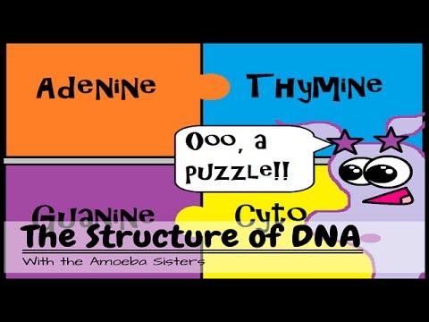
Key Things to Know About DNA Structure

  • DNA is made of nucleotides, each with a sugar (deoxyribose), a phosphate group, and a nitrogenous base
  • The four bases are Adenine (A), Thymine (T), Guanine (G), and Cytosine (C)
  • Base pairing rules: A always pairs with T, and G always pairs with C
  • The two strands run in opposite directions (antiparallel) and are held by hydrogen bonds
  • The outside of the ladder is the sugar-phosphate backbone; the rungs are the base pairs
♻️ Module 4

DNA Replication

Before a cell divides, it must copy its entire DNA so each new cell gets a complete set of genetic instructions.

🎯
SOL Connection: Free nucleotides bond to the template strand (A–T and G–C), forming a complementary strand. The final result is two identical DNA molecules.
βš™οΈ How Replication Works
1
The DNA "Unzips"
An enzyme called helicase breaks the hydrogen bonds between base pairs. The two strands separate β€” like unzipping a zipper β€” creating a replication fork where copying begins.
2
Each Strand Acts as a Template
Each separated strand serves as a template (a pattern) for building a new complementary strand. Free nucleotides floating in the nucleus line up opposite each template base using base-pairing rules: A with T, and G with C.
3
New Strands Are Built
DNA polymerase is the enzyme that connects the free nucleotides together to form the new strand. It "reads" the template and adds the correct matching nucleotide each time.
4
Two Identical DNA Molecules
When replication is complete, there are two identical DNA molecules. Each molecule has one original strand and one newly made strand. This is called semiconservative replication.

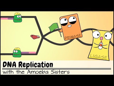
Key Enzymes (Know These!)

  • Helicase β€” unzips the DNA double helix by breaking hydrogen bonds
  • DNA Polymerase β€” builds the new complementary strand using base-pairing rules
πŸ’‘ Semiconservative means each new DNA molecule keeps ("conserves") one original strand and gains one new strand. This ensures faithful copying of genetic information.
βš–οΈ Module 5

DNA vs RNA

DNA and RNA are both nucleic acids, but they have important differences in structure and function.

🎯
SOL Connection: DNA molecules are double-stranded and RNA molecules are single-stranded. One strand of DNA serves as a template to make complementary RNA.
πŸ“Š DNA vs RNA Comparison
Feature 🧬 DNA πŸ”΄ RNA
Full NameDeoxyribonucleic AcidRibonucleic Acid
SugarDeoxyriboseRibose
BasesA, T, G, CA, U (Uracil), G, C
StrandsDouble-strandedSingle-stranded
ShapeDouble helixSingle strand (various shapes)
LocationNucleusNucleus and cytoplasm
FunctionStores genetic informationCarries and uses genetic information to make proteins
🧬 DNA Uses Thymine (T)

In DNA, Thymine (T) pairs with Adenine (A). DNA is very stable because it needs to store information for a long time in the nucleus.

πŸ”΄ RNA Uses Uracil (U)

In RNA, Uracil (U) replaces Thymine and pairs with Adenine (A). RNA is single-stranded and shorter-lived than DNA β€” it is made, used, then broken down.

🧡 Module 6

Types of RNA

There are three main types of RNA involved in protein synthesis. Each has a specific job.

🎯
SOL Connection: Protein synthesis involves mRNA, tRNA, and rRNA working together to build proteins at the ribosome.
πŸ“¨
Messenger RNA
mRNA
Carries the genetic code from DNA in the nucleus out to the ribosome in the cytoplasm. It acts as the "message" or blueprint for building a protein. Each set of three bases on mRNA is called a codon.
🚚
Transfer RNA
tRNA
Brings the correct amino acid to the ribosome. Each tRNA matches a specific mRNA codon using its anticodon. It acts like a delivery truck β€” bringing the right building block at the right time.
🏭
Ribosomal RNA
rRNA
Makes up the structure of the ribosome β€” the organelle where proteins are assembled. The ribosome has two subunits (large and small) that work together to read mRNA and connect amino acids.

Easy Way to Remember

  • mRNA = the Message (carries the code)
  • tRNA = the Truck (delivers amino acids)
  • rRNA = the Ribosome (the factory where it all happens)
✏️ Module 7

Transcription

Transcription is Step 1 of protein synthesis. A section of DNA is copied into mRNA. This happens in the nucleus.

🎯
SOL Connection: One strand of the double-stranded DNA chain serves as a template for the synthesis of a single strand of RNA that is complementary to the DNA strand.
1
RNA Polymerase Binds to DNA
The enzyme RNA polymerase attaches to the beginning of a gene on the DNA strand (a region called the promoter). The DNA double helix unwinds in that region, exposing the template strand.
2
mRNA Is Built Using the DNA Template
RNA polymerase reads the DNA template strand and builds a complementary mRNA strand. Free RNA nucleotides pair with the DNA bases using modified base-pairing rules: A β†’ U, T β†’ A, G β†’ C, C β†’ G. (Notice: wherever the DNA has A, the mRNA gets U β€” not T.)
3
mRNA Is Released and Leaves the Nucleus
When RNA polymerase reaches the end of the gene, the mRNA strand is released. The DNA zips back up. The completed mRNA travels out of the nucleus through a nuclear pore and into the cytoplasm, where translation occurs.
πŸ”€ Transcription Base-Pairing Example
DNA Template:  3'β€” T β€” A β€” C β€” G β€” A β€” T β€” 5'
↓  β†“  β†“  β†“  β†“  β†“
mRNA:         5'β€” A β€” U β€” G β€” C β€” U β€” A β€” 3'

Notice: wherever DNA has A, mRNA gets U β€” not T. This is the key difference between DNA replication and transcription.

πŸ“ Where does transcription happen? In the nucleus. The DNA stays in the nucleus β€” only the mRNA copy travels out to the ribosome.
πŸ”„ Module 8

Translation

Translation is Step 2 of protein synthesis. The ribosome reads the mRNA and builds a protein. This happens in the cytoplasm.

🎯
SOL Connection: Protein synthesis is the process of forming proteins. The mRNA sequence is read three bases at a time (codons) to assemble a chain of amino acids.
1
Ribosome Attaches to mRNA
The ribosome (made of rRNA and protein) attaches to the mRNA strand in the cytoplasm. It finds the start codon β€” the sequence AUG β€” and begins reading there. AUG codes for the amino acid methionine.
2
tRNA Brings Amino Acids
tRNA molecules carry specific amino acids to the ribosome. Each tRNA has an anticodon β€” three bases that match (are complementary to) a codon on the mRNA. When a tRNA anticodon matches the mRNA codon, the correct amino acid is added to the growing protein chain.
3
Amino Acids Are Linked Together
As the ribosome moves along the mRNA one codon at a time, amino acids are linked together by peptide bonds. The chain grows longer with each new amino acid added.
4
Protein Is Released at a Stop Codon
When the ribosome reaches a stop codon (UAA, UAG, or UGA), translation ends. No tRNA matches a stop codon. The completed protein chain is released from the ribosome and folds into its functional shape.
πŸ—οΈ Key Codons to Know
CodonMeaningCodonMeaning
AUGSTART (Methionine)UUU / UUCPhenylalanine
UAASTOPGGU / GGC / GGA / GGGGlycine
UAGSTOPGCU / GCC / GCA / GCGAlanine
UGASTOPAAA / AAGLysine

The complete genetic code has 64 codons for 20 amino acids. Most amino acids have more than one codon β€” this is called redundancy.

Central Dogma of Molecular Biology

  • DNA β†’ mRNA (Transcription, in the nucleus)
  • mRNA β†’ Protein (Translation, at the ribosome in the cytoplasm)
  • DNA is the master copy β€” it stays in the nucleus and never leaves
βš—οΈ Virtual Lab

Interactive Lab

Practice base pairing and codon decoding β€” two core skills on the Virginia Biology SOL.

LAB 1
DNA Complementary Strand Builder
Directions: Click DNA nucleotides to build a template strand (up to 8 bases). Then click Build Complement to see the complementary DNA strand and the mRNA transcript that would be made from this template.
DNA Nucleotides
A
T
G
C
Your Strands
Template:
DNA Comp:
mRNA:
Click nucleotides to build your template strand...
LAB 2
mRNA Codon β†’ Amino Acid Decoder
Directions: Click RNA nucleotides (A, U, G, C) to build an mRNA sequence. Include the start codon AUG to begin translation. Then click Translate to see which amino acids are coded for.
RNA Nucleotides
A
U
G
C
Try: AUG-UUU-GAA-UAA
= Start-Phe-Glu-Stop
mRNA & Translation
mRNA:
Codons:
Protein:
Add RNA nucleotides to begin...
πŸ“ SOL Practice Quiz

Knowledge Check

10 questions written in Virginia Biology SOL style. Answer all questions, then click Submit to see your score and explanations.

🧬

DNA & Protein Synthesis β€” SOL Quiz

10 questions Β· Virginia Biology BIO.3

β€”/10

Your Score

0/10