Unit 2 · Self-Paced Module

Cell Structure
& Function

Master every organelle, membrane process, and cell type aligned to Virginia Biology SOL BIO.3. Move at your own pace — watch, read, practice, and prove it.

BIO.3a Cell theory & organelles
BIO.3b Unicellular vs multicellular
BIO.3c Growth & division
BIO.3d Cell membrane & transport
BIO.3e Cell specialization
Course Progress 0% complete
🎯
What You'll Master
Virginia Biology SOL BIO.3 · ~3 weeks

By the end of this module you'll be able to identify and explain the function of all major organelles, compare prokaryotic and eukaryotic cells, explain how the cell membrane controls what enters and leaves, describe the cell cycle, and explain how cells specialize.

How This Module Works
📋
Self-Paced Flow

Each lesson has 3 parts: Watch a video to build background, Study the interactive slides, then Practice with an assignment or quick check. Mark each lesson complete to track your progress.

Organelles At a Glance
Nucleus
Control center; houses DNA and directs cell activities
Cell Membrane
Selectively permeable barrier; controls what enters/exits
Mitochondria
Powerhouse; converts glucose to ATP via cellular respiration
Ribosome
Site of protein synthesis; found free or on rough ER
Endoplasmic Reticulum
Rough ER: protein processing. Smooth ER: lipid synthesis
Golgi Apparatus
Packages and ships proteins; the cell's "post office"
Lysosome
Digests waste; contains hydrolytic enzymes (animal cells)
Vacuole
Storage; large central vacuole maintains turgor pressure in plants
Chloroplast
Photosynthesis; converts light energy to sugar (plant cells only)
Cell Wall
Rigid support and protection (plants, fungi, bacteria)
Cytoskeleton
Gives shape and moves organelles; made of microtubules & filaments
Centrioles
Organize spindle fibers during cell division (animal cells)
Prokaryote vs Eukaryote
Feature Prokaryote Eukaryote
Membrane-bound nucleus ✗ No ✓ Yes
Membrane-bound organelles ✗ No ✓ Yes
Ribosomes ✓ Small (70S) ✓ Larger (80S)
DNA shape Circular, in cytoplasm Linear, in nucleus
Cell wall Usually present Plants/fungi only
Examples Bacteria, Archaea Plants, Animals, Fungi
📚
5 Lessons · Self-Paced
Complete in any order. Mark each done to update progress.
Lesson 1
1
Cell Theory & Organelle Overview (BIO.3a)
Learn the 3 parts of cell theory, then tour all major organelles and their functions.
▶ Video ◻ Slides ? Quick Check
Click to open →
Lesson 2
2
Prokaryotic vs Eukaryotic Cells (BIO.3a–b)
Compare bacterial cells to plant and animal cells. Unicellular vs multicellular organisms.
▶ Video ◻ Slides ✎ Assignment
Click to open →
Lesson 3
3
Cell Membrane & Transport (BIO.3d)
Phospholipid bilayer, diffusion, osmosis, active vs passive transport, facilitated diffusion.
▶ Video ◻ Slides 🔬 Lab Activity
Click to open →
Lesson 4
4
Cell Growth & Division (BIO.3c)
Cell cycle, interphase, mitosis (PMAT), and cytokinesis. Why cells divide.
▶ Video ◻ Slides ✎ Assignment
Click to open →
Lesson 5
5
Cell Specialization & Differentiation (BIO.3e)
How one fertilized egg becomes trillions of specialized cells. Stem cells, tissues, organs.
▶ Video ◻ Slides ✎ Assignment ? Unit Quiz
Click to open →
Interactive Slide Decks
Click any slide set to view the full presentation
Deck A — Cell Theory & Organelles
01
Cell Theory: The 3 Principles
02
Animal Cell Tour
03
Plant Cell Tour
04
Organelle Functions
05
Prokaryote vs Eukaryote
06
Scale & Microscopy
Deck B — Cell Membrane & Transport
01
Phospholipid Bilayer
02
Passive Transport
03
Osmosis & Tonicity
04
Active Transport
05
Endo & Exocytosis
Deck C — Cell Division
01
The Cell Cycle
02
Mitosis — PMAT
03
Why Cells Divide
04
Cell Cycle & Cancer
Curated Video Lessons
Watch before reading slides for best results. Total: ~75 min.
Lesson 1 — Cell Theory & Organelles
▶ Video 1 · ~15 min
Introduction to Cells — Amoeba Sisters

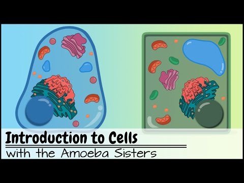
Covers cell theory history, prokaryotic vs eukaryotic comparison, and a tour of organelle functions. Great starting point with clear visuals.

Introduction to Cells thumbnail
Click to watch on YouTube
📝 While watching: List the function of each organelle in your notes. You'll need these for the Assignment 1 diagram.
Lesson 2 — Prokaryote vs Eukaryote
▶ Video 2 · ~12 min
Prokaryotic vs Eukaryotic Cells — Khan Academy

Detailed comparison of bacterial cells and eukaryotic cells, including structural differences and evolutionary relationships.

Prokaryotic vs Eukaryotic thumbnail
Click to watch on YouTube
📝 While watching: Make a T-chart: Prokaryote vs Eukaryote. Note at least 5 differences.
Lesson 3 — Cell Membrane & Transport
▶ Video 3 · ~14 min
Cell Membrane & Transport — Amoeba Sisters

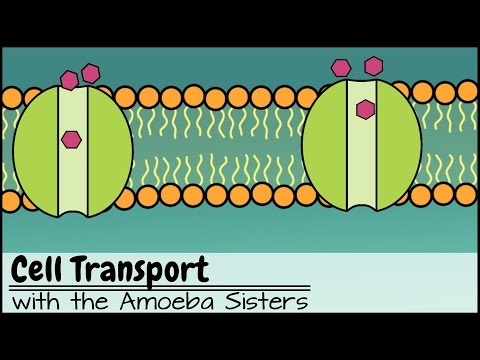
Explains the phospholipid bilayer, selective permeability, diffusion, osmosis, and active transport with memorable analogies.

Cell Membrane Transport thumbnail
Click to watch on YouTube
📝 While watching: Sketch the phospholipid bilayer and label hydrophilic heads and hydrophobic tails.
Lesson 4 — Cell Division / Mitosis
▶ Video 4 · ~11 min
Mitosis: Splitting Up is Complicated — Crash Course Biology

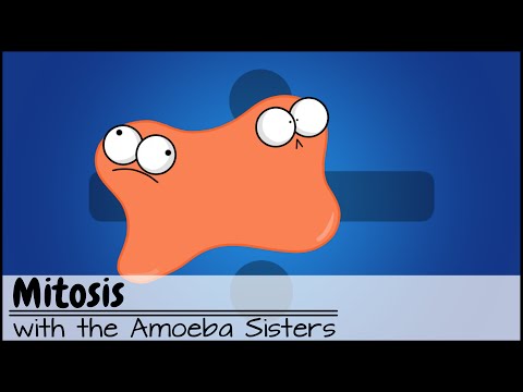
Hank Green walks through the full cell cycle, each phase of mitosis, and why accurate DNA copying matters. Engaging and packed with detail.

Crash Course Mitosis thumbnail
Click to watch on YouTube
📝 While watching: Draw each stage of PMAT with a simple cell diagram showing chromosome position.
Lesson 5 — Cell Specialization
▶ Video 5 · ~10 min
Cell Differentiation — Stated Clearly

Explains how one fertilized egg gives rise to hundreds of cell types through gene expression, including an intro to stem cells.

Cell Differentiation thumbnail
Click to watch on YouTube
📝 While watching: Note 3 examples of specialized cells and explain how their structure fits their function.
4 Assignments · BIO.3 Aligned
Submit to your teacher's preferred platform (Google Classroom, Canvas, etc.)
📐 Assignment 1 — Cell Diagram & Organelle Guide ⏱ After Lesson 1
Create a fully labeled diagram of BOTH a plant cell and an animal cell. For each organelle, write its name, draw it accurately, and describe its function in your own words.
  • Draw a large plant cell (min. half-page). Label: cell wall, cell membrane, nucleus, chloroplast, large central vacuole, mitochondria, ribosomes, ER, Golgi.
  • Draw a large animal cell. Label: cell membrane, nucleus, mitochondria, ribosomes, ER, Golgi, lysosomes, centrioles, small vacuoles.
  • Circle organelles present in BOTH cells in blue. Circle plant-only organelles in green. Circle animal-only organelles in red.
  • Write a 2–3 sentence "function summary" for any 6 organelles of your choice.
  • At the bottom, write: "How is the structure of the mitochondria related to its function?" (3–4 sentences, connect shape to energy production)
🏆 SOL Connection: BIO.3a — Directly tests organelle identification and structure-function relationships.
🔬 Assignment 2 — Osmosis Lab Write-Up ⏱ After Lesson 3
Complete the virtual osmosis lab (using the potato or dialysis tubing simulation) and write a full lab report following the scientific method.
  • Purpose: State the question you are investigating about osmosis and tonicity.
  • Hypothesis: Write an if/then prediction for what will happen to the potato/tubing in hypertonic, hypotonic, and isotonic solutions.
  • Procedure: Summarize the steps in your own words (no copy-paste).
  • Data Table: Record mass/length changes before and after for each solution type.
  • Analysis: Calculate % change in mass. Explain WHY each result occurred using osmosis vocabulary (solute concentration, water potential, tonicity).
  • Conclusion: Was your hypothesis supported? Connect your results to how plant cells maintain turgor pressure and how animal cells respond to different environments (e.g., red blood cells in salt water).
🏆 SOL Connection: BIO.3d — Applies cell membrane transport concepts to experimental data.
🔄 Assignment 3 — Mitosis Sequencing & Analysis ⏱ After Lesson 4
Given microscope images of onion root tip cells, identify each phase of the cell cycle and answer analysis questions.
  • Print or digitally view the provided onion root tip image set (6 images).
  • For each image, identify the cell cycle phase (Interphase, Prophase, Metaphase, Anaphase, Telophase, Cytokinesis).
  • Justify your identification with at least ONE observable feature (e.g., "chromosomes are aligned at the metaphase plate").
  • Count how many cells in your image are in Interphase vs. Mitosis. Calculate the percentage of time spent in each. What does this tell you about which phase takes longest?
  • Short answer (5–6 sentences): Why is it important that mitosis produces genetically identical daughter cells? What would happen if an error occurred during DNA replication?
🏆 SOL Connection: BIO.3c — Cell growth, division, and replication processes.
🧬 Assignment 4 — Cell Specialization Research Card ⏱ After Lesson 5
Choose ONE specialized cell type and create a detailed "Cell Profile Card" that connects structure to function to the broader organism.
  • Choose one: neuron, red blood cell, muscle cell, root hair cell, guard cell, or sperm cell.
  • Draw and label the specialized cell, highlighting features that distinguish it from a "typical" cell.
  • Explain how at least 2 structural adaptations help the cell perform its specific job.
  • Explain how this cell type works with other cell types to support an organ system. (Example: neuron + glial cells + muscle cells = nervous + muscular system communication)
  • Reflection: How does the process of differentiation (all cells having the same DNA but looking different) connect to gene expression? Write 3–4 sentences.
🏆 SOL Connection: BIO.3b & BIO.3e — Multicellular organisms and cell specialization.
?
BIO.3 Practice Quiz
10 questions · SOL-style · Immediate feedback
1. Which organelle is responsible for packaging and shipping proteins to other parts of the cell or outside the cell?
2. A cell is placed in a solution and water moves INTO the cell, causing it to swell. The solution surrounding the cell is best described as —
3. Which of the following is found in plant cells but NOT in animal cells?
4. What is the CORRECT order of the phases of mitosis?
5. A student observes a cell through a microscope and notices it has NO membrane-bound nucleus and NO membrane-bound organelles. This cell is most likely —
6. The movement of molecules from an area of HIGH concentration to LOW concentration WITHOUT using energy is called —
7. Which phase of the cell cycle involves the cell spending the most time growing and preparing for division?
8. A red blood cell and a neuron both have the same DNA, yet they look and function completely differently. This is BEST explained by —
9. The sodium-potassium pump moves 3 Na⁺ ions OUT of the cell and 2 K⁺ ions IN. This is an example of —
10. The cell membrane is described as "selectively permeable." This means —
0/10